[Protein Synthesis, Post-Translational Modification, and Degradation] Adenosine Receptors Control a New Pathway of Fas-associated Death Domain Protein Expression Regulation by Secretion
from Journal of Biological Chemistry current issue by Tourneur, L., Mistou, S., Schmitt, A., Chiocchia, G.
FADD is the key adaptor transmitting the apoptotic signal mediated by death receptors. We have previously shown that FADD protein expression could be lost in vivo in cancerous cells, in mice and humans, and be used as prognostic factor. Furthermore, loss of FADD could contribute to tumor progression and aggressiveness. However, the mechanism accounting for the loss of FADD was unknown. Using in vitro-cultured mouse organ models, we demonstrated that loss of FADD occurred through a new regulatory pathway of FADD expression by secretion. The secretion of FADD is an active release following shedding of microvesicles derived from the plasma membrane. In our experimental settings, this phenomenon was restricted to 6 of 12 FADD-expressing organs. This process is calcium- and adenosine-dependent. Moreover, we identified the two receptors with low affinity to adenosine, namely A2B and A3 adenosine receptors, as regulators of the FADD secretion process. Furthermore, we showed that modulating A3 adenosine receptor can convert a nonsecreting organ into a FADD-secreting one. Finally, we reported that mouse FADD release occurred in vivo during tumor disease. These results demonstrate the existence of a new localization site (in microvesicles) and regulatory mechanism (by secretion) of the FADD protein, and the implication of adenosine receptors in this process. These data open a new field of investigation consisting of the possibility to regulate FADD expression via the modulation of adenosine receptors, which constitutes a therapeutic target in diseases in which FADD-mediated signaling is impaired.
Monday, June 30, 2008


Friday, June 27, 2008
SATB1 paper importante
Nature Clinical Practice Oncology (2008) 5, 364
doi:10.1038/ncponc1141 
SATB1 expression generates gene-expression profiles that promote breast tumor metastasis
Original article
Han HJ et al. (2008) SATB1 reprogrammes gene expression to promote breast tumour growth and metastasis. Nature 452: 187–193 PubMed
Gene-expression analyses of human breast cancer cell lines that are associated with a poor outcome have revealed characteristic expression patterns that can predict metastasis (or metastatic risk), but how such gene-expression profiles confer metastatic potential is unclear. Han et al. have described a genome organizer protein—SATB1—that when expressed in breast cancer cells can establish a pattern of gene expression that is consistent with invasive behavior.
Initial investigations involving breast epithelial cell lines and breast-tumor specimens revealed that SATB1 is expressed only in metastatic cancer cell lines. To determine the prognostic significance of SATB1 expression, tissue microarrays comprising 1,318 breast cancer samples were scored on the basis of expression levels of SATB1 protein in tumor cell nuclei. Kaplan–Meier analysis of data from 985 patients with ductal breast carcinomas revealed a correlation between higher levels of SATB1 expression and shorter overall patient survival (P <0.001).>in vitro proliferation, restored anchorage-dependent growth and breast-like acinar polarity, and inhibited tumor growth and metastasis in vivo. Consistent with these findings, ectopic expression of SATB1 in a nonmetastatic cell line was sufficient to confer invasive activity in vivo. Global gene-expression profiling showed that expression of SATB1 altered the expression patterns of over 1,000 genes. Further analysis revealed that SATB1 modified the epigenetic status of all SATB1-dependent genes analyzed.
The authors conclude that SATB1 establishes gene-expression profiles that promote tumor growth and metastasis.
Sunday, June 22, 2008
La caspasa 9 debe procesarse en la apoptosis?
COMMENTARY
Caspase-9 cleavage, do you need it?
Davina TWIDDY and Kelvin CAIN1
MRC Toxicology Unit, Hodgkin Building, University of Leicester, Lancaster Rd, Leicester LE1 9HN, U.K.
Caspase-9, which is activated by association with the Apaf-1
(apoptotic protease-activating factor-1) apoptosome complex,
cleaves and activates the downstream effector caspases-3 and -7,
thereby executing the caspase-cascade and cell-death programme.
Although caspase-9 does not need to be cleaved to be active, apoptotic
cell death is always accompanied by autocatalytic cleavage
and by further downstream effector caspase-dependent cleavage
of caspase-9. In this issue of the Biochemical Journal,Denault
and co-workers evaluate the role of caspase-3-dependent cleavage
of caspase-9 and conclude that this mechanism mainly serves
to enhance apoptosis by alleviating XIAP (X-linked inhibitor of
apoptosis) inhibition of the apical caspase.
Biochem. J. (2007) 405, e1–e2 (Printed in Great Britain) doi:10.1042/BJ20070617
Promotores y splicing
Alternative Promoters Influence Alternative Splicing at the Genomic Level
Abstract
Background
More and more experiments have shown that transcription and mRNA processing are not two independent events but are tightly coupled to each other. Both promoter and transcription rate were found to influence alternative splicing. More than half of human genes have alternative promoters, but it is still not clear why there are so many alternative promoters and what their biological roles are.
Methodology/Principal Findings
In this study, we explored whether there is a functional correlation between alternative promoters and alternative splicing by a genome-wide analysis of human and mouse genes. We constructed a large data set of genes with alternative promoter and alternative splicing annotations. By analyzing these genes, we showed that genes with alternative promoters tended to demonstrate alternative splicing compare to genes with single promoter, and, genes with more alternative promoters tend to have more alternative splicing variants. Furthermore, transcripts from different alternative promoters tended to splice differently.
Conclusions/Significance
Thus at the genomic level, alternative promoters are positively correlated with alternative splicing.
ver articulo en PLOS one
Monday, June 16, 2008
Salud en Mexico. PLOS Medicine
Characterizing the Epidemiological Transition in Mexico: National and Subnational Burden of Diseases, Injuries, and Risk Factors
1 Harvard School of Public Health, Boston, Massachusetts, United States of America, 2 Harvard Initiative for Global Health, Cambridge, Massachusetts, United States of America, 3 World Health Organization, Geneva, Switzerland, 4 Pennsylvania State University, University Park, Pennsylvania, United States of America, 5 Instituto Nacional de Salud Pública, Cuernavaca, Mexico
Background
Rates of diseases and injuries and the effects of their risk factors can have substantial subnational heterogeneity, especially in middle-income countries like Mexico. Subnational analysis of the burden of diseases, injuries, and risk factors can improve characterization of the epidemiological transition and identify policy priorities.
Methods and Findings
We estimated deaths and loss of healthy life years (measured in disability-adjusted life years [DALYs]) in 2004 from a comprehensive list of diseases and injuries, and 16 major risk factors, by sex and age for Mexico and its states. Data sources included the vital statistics, national censuses, health examination surveys, and published epidemiological studies. Mortality statistics were adjusted for underreporting, misreporting of age at death, and for misclassification and incomparability of cause-of-death assignment. Nationally, noncommunicable diseases caused 75% of total deaths and 68% of total DALYs, with another 14% of deaths and 18% of DALYs caused by undernutrition and communicable, maternal, and perinatal diseases. The leading causes of death were ischemic heart disease, diabetes mellitus, cerebrovascular disease, liver cirrhosis, and road traffic injuries. High body mass index, high blood glucose, and alcohol use were the leading risk factors for disease burden, causing 5.1%, 5.0%, and 7.3% of total burden of disease, respectively. Mexico City had the lowest mortality rates (4.2 per 1,000) and the Southern region the highest (5.0 per 1,000); under-five mortality in the Southern region was nearly twice that of Mexico City. In the Southern region undernutrition and communicable, maternal, and perinatal diseases caused 23% of DALYs; in Chiapas, they caused 29% of DALYs. At the same time, the absolute rates of noncommunicable disease and injury burdens were highest in the Southern region (105 DALYs per 1,000 population versus 97 nationally for noncommunicable diseases; 22 versus 19 for injuries).
Conclusions
Mexico is at an advanced stage in the epidemiologic transition, with the majority of the disease and injury burden from noncommunicable diseases. A unique characteristic of the epidemiological transition in Mexico is that overweight and obesity, high blood glucose, and alcohol use are responsible for larger burden of disease than other noncommunicable disease risks such as tobacco smoking. The Southern region is least advanced in the epidemiological transition and suffers from the largest burden of ill health in all disease and injury groups.
Thursday, June 12, 2008
Creacion de nuevos genes
Research Highlight
Nature Reviews Genetics 9, 415 (June 2008) | doi:10.1038/nrg2394
Evolution: A gene is born
Tanita Casci

There are various ways to explain how new genes can come about — exon shuffling, gene duplication and retroposition being just a few. But how are truly novel genes born out of a sequence that was previously non-coding? Using a comparative genomics approach, a research group has identified and characterized the function of a novel ORF in Saccharomyces cerevisiae, and has shown that it might contribute to the evolution of this yeast species.
The mechanism by which a gene is created from scratch receives relatively little attention, despite reports from sequencing projects and comparative analyses that many de novo genes exist in some bacterial and animal lineages. To investigate the process of gene creation, the authors concentrated on BSC4, a species-specific gene that was pulled out of genome comparisons among Saccharomyces species. Although the gene has no homologue in other fungal species, the region around BSC4 is syntenic across Saccharomyces species — suggesting that it probably did not jump into its current position on the S. cerevisiae genome by horizontal gene transfer, and allowing the birth of the BSC4 ancestral sequence to be dated to over 100 million years ago.
How do we know that BSC4 is a real protein-coding gene? Most criteria would suggest that it is: the sequence — an ORF of 132 amino acids — is fixed in S. cerevisiae strains, and RT-PCR experiments suggest that the ORF can be transcribed. Proteomics analyses support the existence of 29 peptides associated with this locus, and the BCS4 sequence seems to be under purifying selection. It is not clear what BSC4 actually does, but the fact that it is synthetically lethal with mutations in two DNA-damage repair genes makes it likely that it acts in this pathway.
This paper provides the first evidence that new genes can arise from a non-coding region that is initially transcribed and then subsequently obtains an ORF via mutation. The fact that BSC4 expression is upregulated during the stationary phase of the cell cycle, when DNA repair is most pressing, supports the idea that not only is this new gene real, it is also selectively advantageous.
Tuesday, May 27, 2008
Idea?
Este estudio utiliza Chip para mapear sitios y genes ubiquitinados en el genoma
http://www.signaling-gateway.org/update/updates/200805/nmeth0508-380.html
Podria hacerse algo parecido con los factores de splicing? Quiza para mapear regulacion trans de splicing despues de estimulos especificos?
http://www.signaling-gateway.org/update/updates/200805/nmeth0508-380.html
Podria hacerse algo parecido con los factores de splicing? Quiza para mapear regulacion trans de splicing despues de estimulos especificos?
Friday, May 23, 2008
Nuevo mecanismo? Activacion por 5 UTRs.
PLOS Medicine
A Novel Tumor-Promoting Function Residing in the 5′ Non-coding Region of vascular endothelial growth factor mRNA
Background
Vascular endothelial growth factor-A (VEGF) is one of the key regulators of tumor development, hence it is considered to be an important therapeutic target for cancer treatment. However, clinical trials have suggested that anti-VEGF monotherapy was less effective than standard chemotherapy. On the basis of the evidence, we hypothesized that vegf mRNA may have unrecognized function(s) in cancer cells.
Methods and Findings
Knockdown of VEGF with vegf-targeting small-interfering (si) RNAs increased susceptibility of human colon cancer cell line (HCT116) to apoptosis caused with 5-fluorouracil, etoposide, or doxorubicin. Recombinant human VEGF165 did not completely inhibit this apoptosis. Conversely, overexpression of VEGF165 increased resistance to anti-cancer drug-induced apoptosis, while an anti-VEGF165-neutralizing antibody did not completely block the resistance. We prepared plasmids encoding full-length vegf mRNA with mutation of signal sequence, vegf mRNAs lacking untranslated regions (UTRs), or mutated 5′UTRs. Using these plasmids, we revealed that the 5′UTR of vegf mRNA possessed anti-apoptotic activity. The 5′UTR-mediated activity was not affected by a protein synthesis inhibitor, cycloheximide. We established HCT116 clones stably expressing either the vegf 5′UTR or the mutated 5′UTR. The clones expressing the 5′UTR, but not the mutated one, showed increased anchorage-independent growth in vitro and formed progressive tumors when implanted in athymic nude mice. Microarray and quantitative real-time PCR analyses indicated that the vegf 5′UTR-expressing tumors had up-regulated anti-apoptotic genes, multidrug-resistant genes, and growth-promoting genes, while pro-apoptotic genes were down-regulated. Notably, expression of signal transducers and activators of transcription 1 (STAT1) was markedly repressed in the 5′UTR-expressing tumors, resulting in down-regulation of a STAT1-responsive cluster of genes (43 genes). As a result, the tumors did not respond to interferon (IFN)α therapy at all. We showed that stable silencing of endogenous vegf mRNA in HCT116 cells enhanced both STAT1 expression and IFNα responses.
Conclusions
These findings suggest that cancer cells have a survival system that is regulated by vegf mRNA and imply that both vegf mRNA and its protein may synergistically promote the malignancy of tumor cells. Therefore, combination of anti-vegf transcript strategies, such as siRNA-based gene silencing, with anti-VEGF antibody treatment may improve anti-cancer therapies that target VEGF.
Monday, April 28, 2008
Valdría la pena probar?
Pimp Your Plasmid Growth Medium
 I often wonder why it is that molecular biology researchers stubbornly refuse to change 40 year old methods that, while they work, are not as good as newer, faster and cheaper methods out there.
I suppose rational scientists have often irrational superstitions.
One example of an old method that could be improved is the growth media used for plasmid preparation.
The majority of us, throughout our university careers, have used either SOC, LB or TB, for recombinant plasmid propagation, typically in E. coli. LB or Luria-Bertani broth has been in use for almost 60 years or thereabouts, while SOC has certainly been in use for 2 decades.
But by adding in a few more ingredients or being more economical on others (especially yeast extract and tryptone) that you could get a higher plasmid yield, quicker and with less money.
To counter the naysayers, nobody wants to make very complex with 15 ingredients requiring filter sterilisation, as this obviously defeats the object of economy of time and budget. Indeed, there are trade-offs between optimising for biomass, plasmid yield, quality, stability and cost with the difference between protein production and plasmid production being that plasmid production requires only cell growth, division, and plasmid stability.
The good news is that Michael Danquah and Gareth Forde from Monash University down-under have devised a stoichiometrically optimised medium for plasmid production. PDM, supposedly yields under the conditions they tested, twice the amount of plasmid in both volumetric and specific yields compared to TB , LB is left in the dust. Better yet, because it uses less tryptone and yeast extract, the cost per mg of DNA is roughly one quarter compared to LB.
The recipes for LB, TB, SOC and PDM are shown below. If you decide to break with tradition and give PDM a go, be sure to tell us how it goes.
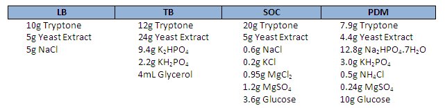
Note – Autoclave glucose, KH2PO4 and Na2HPO4 separately
Monday, April 14, 2008
Peligros de la contaminacion con micoplasma
Oncogene advance online publication 14 April 2008; doi: 10.1038/onc.2008.103
Mycoplasma infection suppresses p53, activates NF- B and cooperates with oncogenic Ras in rodent fibroblast transformation
Prokaryotes of the genus Mycoplasma are the smallest cellular organisms that persist as obligate extracellular parasites. Although mycoplasma infection is known to be associated with chromosomal instability and can promote malignant transformation, the mechanisms underlying these phenomena remain unknown. Since persistence of many cellular parasites requires suppression of apoptosis in host cells, we tested the effect of mycoplasma infection on the activity of the p53 and nuclear factor (NF)-
 B pathways, major mechanisms controlling programmed cell death. To monitor the activity of p53 and NF-B in mycoplasma-infected cells, we used a panel of reporter cell lines expressing the bacterial
 -galactosidase gene under the control of p53- or NF-B-responsive promoters. Cells incubated with media conditioned with different species of mycoplasma showed constitutive activation of NF-B and reduced activation of p53, common characteristics of the majority of human tumor cells, with M. arginini having the strongest effect among the species tested. Moreover, mycoplasma infection reduced the expression level and inducibility of an endogenous p53-responsive gene, p21waf1, and inhibited apoptosis induced by genotoxic stress. Infection with M. arginini made rat and mouse embryo fibroblasts susceptible to transformation with oncogenic H-Ras, whereas mycoplasma-free cells underwent irreversible p53-dependent growth arrest. Mycoplasma infection was as effective as shRNA-mediated knockdown of p53 expression in making rodent fibroblasts permissive to Ras-induced transformation. These observations indicate that mycoplasma infection plays the role of a p53-suppressing oncogene that cooperates with Ras in cell transformation and suggest that the carcinogenic and mutagenic effects of mycoplasma might be due to inhibition of p53 tumor suppressor function by this common human parasite.
Tuesday, April 08, 2008
Wednesday, April 02, 2008
Quick and dirty
Identificacion de plasmidos con inserto en clonaciones
Reagents:
2x Lysis Buffer:
20% w/v Sucrose
200mM NaOH
120mM KCl
10mM EDTA
0.5% SDS
a pinch of Bromophenol blue
store at -20 degC
Procedure:
There are two ways of performing this screen. If the chances of you having a clones depend on the number of colonies you screen i.e. your “luckiness” factor is abysmal, then obviously you would have a screen a large number of colonies. Then go ahead with the 96 well plate method. If you are the type who usually strikes on the clone in a screen of say 10 to 20 colonies then you could use Eppendorf tubes.
1. Dilute lysis buffer to 1x, at room temperature.
2. Pick the colonies from the transformant plate “patch” to a master plate and transfer remaining to 30 microL of lysis buffer (in Eppendorfs or a 96 well plate)
3. Incubate at 37 degC for 5 to 7 minutes
4. Chill on ice for 5min
5. Spin at maximum speed for 10 minutes ( you would need a compatible centrifuge to spin 96 well plates - which should be sealed with parafilm)
6. Load 10 to 15 microL on the gel (Caution: When trying to load from the lysate make sure you only pick up the supernatant. The gooey mass below (caused by genomic DNA) will stick to your piptte causing pipetting difficulties as well cause the sample to jump out of the agarose lanes. If this happens spin down again for a few minutes and load)
7. Difference in electrophoretic mobility will help you narrow down your clones for mini preps.
http://bitesizebio.com/2008/04/02/quick-and-dirty-screening-for-cloned-inserts/
Subscribe to:
Posts (Atom)